Technical advantages
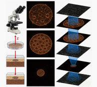
Z-axis layer scan: Uses patented lenses capable of scanning depths over 10mm and supports at least 100 layers, enabling automated scanning to balance deep sample detail capture and high-throughput experimental efficiency requirements.
Spatial Positioning: Combined with automated imaging technology, it can accurately locate various organoid samples in different culture vectors, and adapts to a stable culture environment to dynamically track specific locations within the same batch over the long term, meeting the needs of full-process localization and growth monitoring needs.
Application scenarios
Brightfield, contrast, and fluorescence observation of 2D cells/3D organoids/tissues
Real-time 3D noise reduction, depth fusion, large-format stitching, and layered stitching
Single field of view, 3×3 field of view, 5×5 field of view
Tumor research, drug screening, personalized medicine, gene editing validation
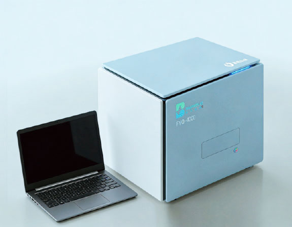
Equipment advantages
Fully automatic high-throughput scanning
Supports mainstream brand petri dishes, honeycomb chips, and coral chips, with laminated scan splicing.
Deep learning algorithms
Patented AI model identifies organoid morphology/volume/fluorescence intensity.
Multidimensional data analysis
Automatically generates growth curves, drug susceptibility scores, and phenotype classification reports.
Online consultation